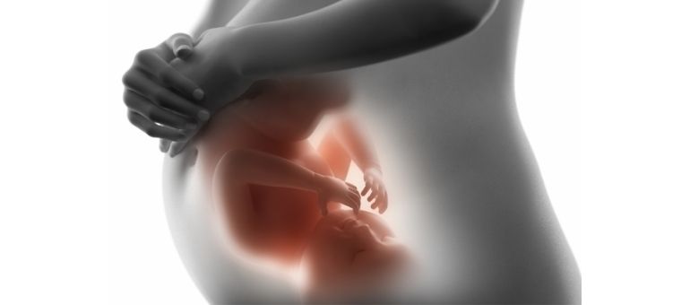
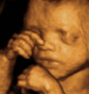
4D (FOUR-DIMENSIONAL), COLOR, DETAILED PREGNANCY ULTRASOUND
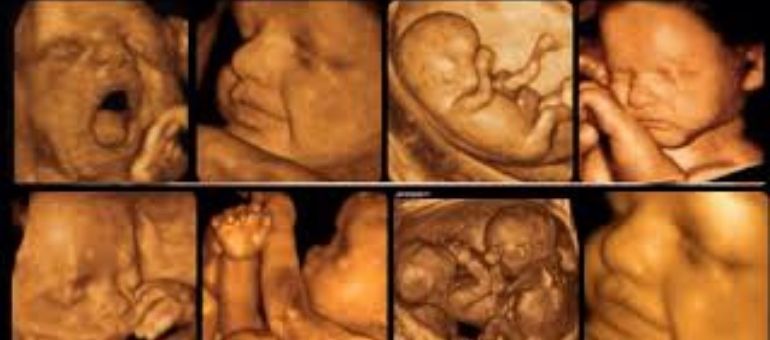
The photographic 3D (three-dimensional) image of a fetus was first made possible in the mid-90s with 4D (four-dimensional) Kretz technology. This technology added the factor of time to photographic images, making it possible to take videos in addition to the static photos we use today. This ultrasound examination, performed between the 18th and 23rd weeks of pregnancy, was previously known as ‘second-level’ or ‘color ultrasound.’ Today, doctors and patients mostly use the names ‘4D (four-dimensional)‘ and/or ‘Detailed Ultrasound.’ Regardless of which name is preferred, they all refer to the same examination, and the main goal is fetal anomaly screening.
We encounter 3 groups of expectant mothers in this examination:
The first group includes expectant mothers with a risk of fetal anomalies or disease/high-risk pregnancies:
- Consanguineous marriage
- Suspicious fetal anomalies or findings detected in previous ultrasound examinations
- A history of a fetus with anomalies in a previous pregnancy
- A maternal, paternal, or familial history of anomalies
- Twin pregnancy
- IVF/in vitro fertilization pregnancy
- Pregnancy diabetes
- Maternal age over 35
- Those with positive results on double or triple tests
- Fetal developmental disorders
- Decreased or increased amniotic fluid
- A history of maternal infections like Toxoplasmosis, CMV, or the use of drugs, alcohol, or narcotics
The second group includes mothers whose pregnancy is progressing normally but who are curious about their baby’s health inside and want to know that everything is okay.
The third group includes those who are simply curious about the 4D (four-dimensional) examination and images of the fetus.
The ultrasound examination for all 3 groups of expectant mothers is successfully performed in our center.
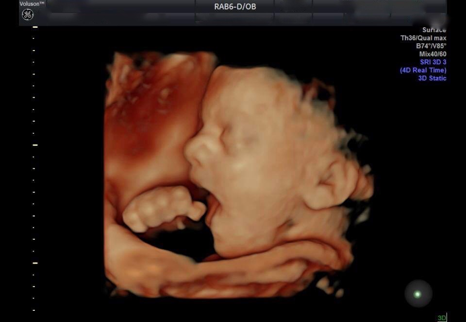
Our main goal with 4D (four-dimensional), detailed ultrasound is to screen for fetal organ and chromosomal anomalies during the pregnancy process. These anomalies can be major/significant (2-3%) or minor/less significant (15%). Especially with the advances in ultrasound technology and equipment, the accumulation of knowledge, and the increasing experience of physicians over the last 10 years, it is possible to diagnose up to 75% of major anomalies before birth. If an anomaly is detected, this examination allows us to provide the family with options regarding the continuation, monitoring, termination, or, in rare cases, treatment of the pregnancy. An important point is that some fetal anomalies can only be diagnosed in the later weeks of pregnancy. Meanwhile, some anomalies do not show any signs throughout the entire pregnancy process and can only be diagnosed after birth. It is also important to note that finding no abnormal signs during the ultrasound examination does not guarantee that the baby is absolutely normal. The expectant mother should keep this in mind.
During the 4D (four-dimensional), detailed ultrasound, we also evaluate uterine shape anomalies and wall problems, cervical length, the number of fetuses, heartbeat and viability, the amount of amniotic fluid, fetal development, and blood flow in the baby and uterus with a color Doppler ultrasound scan. The risks of pregnancy hypertension, placental insufficiency, and pre-eclampsia are also identified. And finally, the expectant mother receives a report, a CD with the information, images, and mini-videos obtained during the examination.