Myomas are benign, non-cancerous tumors that originate from the muscular layer of the uterus.
WHAT IS A MYOMA (FIBROID)?
Myomas are benign, non-cancerous tumors that originate from the muscular layer of the uterus. They are also known as fibroids, leiomyomas, or fibromyomas. These tumors can range in size from 1 to 20 cm and are a condition that can be seen in approximately 25-50% of all women, and in one out of every three women over the age of 35. They can rarely be seen starting from age 15. Patients may have a single myoma or multiple myomas of varying sizes located in different areas. It is difficult to predict whether myomas will appear alone or in groups. They can remain very small for a long time, suddenly grow rapidly, or grow slowly over many years.
WHAT ARE THE CAUSES OF MYOMAS (FIBROIDS)?
Although myomas are very common, little is known about what causes them. Estrogen, known as the female hormone, causes myomas to grow. For this reason, myomas typically grow by about 2-3 times during a woman’s reproductive years and in pregnancy. During menopause, myomas usually shrink because estrogen levels decrease.
WHAT ARE THE TYPES OF MYOMAS (FIBROIDS)?
Myomas are divided into 3 groups based on their location in the uterine tissue:
SUBMUCOSAL MYOMAS
These are myomas that grow toward the inner cavity of the uterus. Although they are the least common type of myoma, they are the ones that cause the most bleeding problems. They put pressure on the endometrium, the inner lining of the uterus, causing damage. They lead to an increase in the amount of menstrual bleeding, prolonged bleeding, heavy, clotted bleeding, bleeding between periods, and serious anemia as a result. Sometimes they can grow out from the cervix into the vagina. In these cases, complaints of pain and bleeding during sexual intercourse may occur.
INTRAMURAL MYOMAS
These are myomas located in the middle of the uterine wall, within the muscle tissue. They are the most common type of myoma. Like submucosal myomas, they can cause increased menstrual bleeding, prolonged bleeding, and related anemia. Additionally, these myomas can cause the uterus to grow, leading to abdominal pain and a feeling of fullness. Large myomas and the resulting enlarged uterus can also put pressure on surrounding organs. Pressure on the intestines can lead to an inability to have bowel movements and chronic constipation. Pressure on the bladder can cause problems like difficulty urinating or, conversely, frequent urination.
SUBSEROSAL MYOMAS
These are myomas that grow from the outer surface of the uterus into the abdominal cavity. They do not typically cause bleeding problems. Depending on the size of the myoma, pressure symptoms are more prominent. They often cause symptoms of pressure, such as abdominal pain, abdominal cramps, back pain, a feeling of fullness, difficulty urinating or frequent urination, and constipation.
WHAT ARE THE SYMPTOMS OF MYOMAS (FIBROIDS)?
Most myomas do not cause any symptoms and do not require treatment. However, about 10-20% of all myomas can cause some symptoms depending on their location:
- Menstrual changes: Irregular periods, longer or more frequent periods, vaginal bleeding between periods, heavier bleeding than normal, heavy and clotted vaginal bleeding.
- Pain: Menstrual cramps, abdominal pain and a feeling of fullness, abdominal cramps, back, lower back, rectal, and leg pain (often a dull, heavy, and aching pain), pain during sexual intercourse.
- Pressure: Difficulty urinating or frequent urination (due to the myomas putting pressure on the bladder), inability to have a bowel movement and chronic constipation (due to the myomas putting pressure on the bowel system).
- Infertility: Especially submucosal myomas that grow toward the inner cavity of the uterus can prevent a pregnancy from implanting.
- Growth in the uterus and abdomen, abdominal swelling.
While most myomas do not cause problems, complications can rarely develop. Myomas attached to the uterine surface by a stalk can become twisted (the myoma twists on its axis, cutting off its blood supply). In these cases, pain, nausea, or fever may occur. Myomas can become infected. In very rare cases, a rapidly growing myoma and other symptoms may indicate a cancerous transformation.
MYOMAS (FIBROIDS) AND PREGNANCY
Myomas can be seen in some pregnant women and usually do not cause any problems. During pregnancy, the size of myomas increases with the increased blood flow to the uterus and the rise in estrogen hormones. The growth of myomas can cause a feeling of pressure and pain. In the majority of patients, the size of the myomas decreases after pregnancy. Myomas can increase the risk of:
- Miscarriage (a pregnancy ending before 20 weeks).
- Premature birth.
- Breech birth (the baby is in a position other than head-down).
- More blood loss during and/or after childbirth.
Rarely, a large myoma can block the cervix or prevent the baby from passing through the birth canal. In these cases, delivery is usually done by Cesarean section. Myomas do not usually need to be treated during pregnancy. A myomectomy (myoma removal surgery) can be performed on a pregnant woman only in very rare cases, or a myomectomy may be needed during a Cesarean section. Sometimes, a pregnant woman with myomas may need to rest or be hospitalized for a period due to pain, bleeding, or the threat of premature birth.
HOW ARE MYOMAS (FIBROIDS) DIAGNOSED?
Most myomas can be detected with a gynecological exam and ultrasound. Sometimes, additional diagnostic methods may be used:
- Hysteroscopy: This uses a thin instrument called a hysteroscope with a camera on the end to see inside the uterus. The instrument is inserted through the vagina and into the cervix. This allows for the visualization of myomas inside the uterine cavity.
- Hysterosalpingography (HSG): This is a special X-ray that can detect abnormal changes in the shape and size of the uterus and fallopian tubes.
- Saline Infusion Sonography (SIS): This involves injecting a saline solution into the uterus and examining it with an ultrasound. This can help distinguish between myomas and polyps inside the uterus.
- Laparoscopy: Myomas on the outer surface of the uterus can be detected with a camera inserted through a small skin incision in or just below the belly button.
- Magnetic Resonance Imaging (MRI) and Computed Tomography (CT): These are rarely necessary. Sometimes, myomas can be incidentally detected when these scans are used to investigate another medical problem or symptom.
WHAT ARE THE TREATMENT OPTIONS FOR MYOMAS (FIBROIDS)?
Small myomas that do not cause any symptoms and those found in women approaching menopause do not often require treatment. There is no need to limit sexual activity unless the myomas cause pain during intercourse. For myomas that do require treatment, the options vary depending on factors such as the patient’s age, desire to have children, and the myoma’s location, number, size, and growth rate. Myomas can be treated with medication or surgery.
1. MEDICATIONS
Medications like gonadotropin-releasing hormone (GnRH) agonists can be used to temporarily shrink the size of myomas and control bleeding in preparation for surgery.
2. SURGERY
MYOMECTOMY
Myomectomy is the surgical removal of only the myomas, leaving the uterus in place. With this surgical method, the patient’s uterus is preserved, so there is a chance to have children. Patients who have a myomectomy can still have a natural vaginal birth if they become pregnant after the surgery, but a C-section may be necessary. During myoma removal, especially in patients who wish to have children, it is important to remove the myomas without damaging the endometrium lining the uterine wall and to not compromise the integrity of the uterus. It should be noted that myomas can recur over time after the surgery. Myomectomy can be performed in several ways. The surgical technique used depends on the location, number, and size of the myomas.
Submucosal myomas that grow toward the inner cavity of the uterus can be removed with hysteroscopic surgery performed through the vagina without any abdominal incision. While this method cannot remove myomas within the uterine wall or on the outer surface, it can often control the bleeding caused by the myomas. After a hysteroscopy, it is usually not necessary to stay in the hospital overnight. Myomas that grow within the uterine wall or toward the outside of the uterus can be removed with laparoscopic (minimally invasive) or laparotomy (open) surgery. For laparotomy, an incision is made in the abdomen, and the myomas are then removed through this incision. Myomas can also be removed via a laparoscope, which is used to see inside the abdomen.
Myoma surgery can also be performed on virgin patients. The hymen is not damaged when laparoscopic (minimally invasive) or laparotomy (open) surgery is performed.
HYSTERECTOMY
Hysterectomy is the treatment of a patient by surgically removing the entire uterus along with the myoma, via laparoscopic (minimally invasive), laparotomy (open), or vaginal approaches. The surgical method is determined based on the size of the uterus and the size, number, and location of the myomas. After a hysterectomy, a patient can no longer have children.
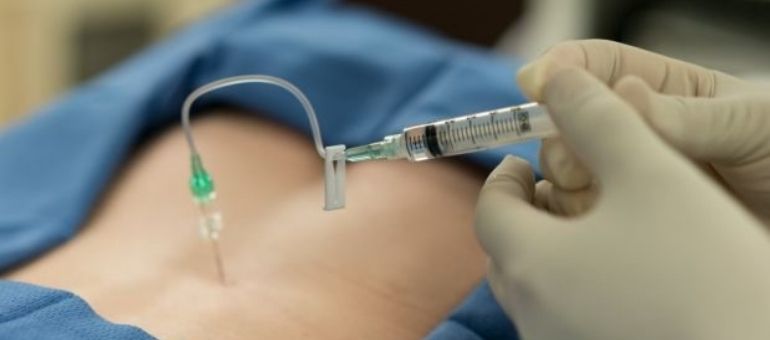
UTERINE ARTERY EMBOLIZATION
With this method, the blood vessels going to the uterus are blocked in a controlled manner. The blood flow that allows myomas to grow is stopped. This causes the myomas to shrink.